he
anatomical blind spot
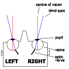
The
area where the optic nerve connects to the retina in the back of each eye is
known as the optic disk. There is a total absence of cones and rods in this
area, and, consequently, each eye is completely blind in this spot. Under
normal binocular vision conditions this is not a problem, because an object
cannot be in the blind spot of both eyes at the same time. On the other
hand, where the field of vision of one eye is obstructed by an object
(windshield post), a visual target (another aircraft) could fall in the
blind spot of the other eye and remain undetected.
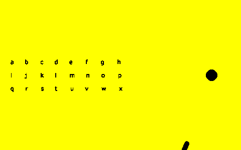
In order to find the blind spot of the right eye, it is necessary to close
the left eye, focus the right eye on a single point, and see if anything
vanishes from vision some 20 degrees right of this point. The following diagram
has a set of characters on the left hand side, and black circle on the right.
Keeping your head motionless, with the right eye about 3 or 4 times as far from
the page as the length of the red line, look at each character in turn, until
the black circle vanishes.

With increasing age, the blind spot enlarges. You may find that the black
circle disappears when several of the characters are looked at. The size and
shape of the blind spot can be found if a large enough grid of characters is
used.
The same test can be done for the left eye. Close the right eye, and look at
each character until the black circle disappears.

Note that when the black circle vanishes, you see only a white background
where the circle was. What happens if the background colour is different? Say,
yellow.

The blind spot appears as yellow. This is interesting, because it means that,
although my eye can't detect anything in the blind spot, something knows that it
is surrounded by yellow, and has guessed that what is in the blind spot is
probably yellow. Smart!
How smart? If a thick horizontal line is drawn through the blind spot, what
happens then?

The answer, it seems, is that if the line passes right through the blind
spot, whatever is making shrewd guesses about colours is also able to work out
that a line going in one side and coming out the other probably continues
through the middle. The black circle disappears, but the line remains.
So what happens when a pen or pencil is pushed into the blind spot? It seems
that as the tip enters the blind spot, the pencil appears truncated, as if it
were vanishing into something (which, after all, it is). But when the tip
emerges at the other side, the visual processing system fills in the missing
part between. The following animation mimics pushing a pencil into the blind
spot.
The first conclusion drawn from this little experiment is that, although each
eye has a blind spot, some sort of intelligence is used to give this area not
only a likely colour, but also to fill in lines that pass through the blind spot
- rather than just have a fuzzy grey area. The net result is that, with one eye
closed, it isn't immediately obvious where the blind spot is, because it has
been given a suitable colour, and even pattern, based on what is adjacent to it.
The second conclusion drawn is that what we see is not just what has appeared
on the retina, but is an image that has been reprocessed, tidied up. And if the
human visual cortex is able to tidy up the blind spot, then it may well be that
the same is being done for the entire visual field - that what we get to 'see'
is not what appears on the retina, like a photograph, but instead something
which has a whole bunch of special effects added.
If so, then we can't trust our eyes. We're being given doctored information,
massaged figures. The world that we see is not something out there, but a world
that we invent. The world I see is my idea.
The Night Blind Spot
The
"Night Blind Spot" appears under conditions of low ambient illumination due
to the absence of rods in the fovea, and involves an area 5 to 10 degrees
wide in the centre of the visual field. Therefore, if an object is viewed
directly at night, it may go undetected or it may fade away after initial
detection due to the night blind spot.
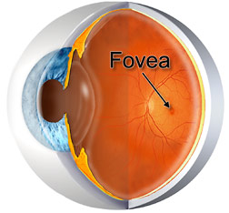
The Fovea
The fovea is the small depression located in the exact
centre of the
macula that contains a high concentration of cones but no rods, and this is
where our vision is most sharp. While the normal field of vision for each
eye is about 135 degrees vertically and about 160 degrees horizontally, only
the fovea has the ability to perceive and send clear, sharply focused visual
images to the brain. This foveal field of vision represents a small conical
area of only about 1 degree. To fully appreciate how small a one-degree
field is, and to demonstrate foveal field, take a quarter from your pocket
and tape it to a flat piece of glass, such as a window. Now back off 4 1/2
feet from the mounted quarter and close one eye. The area of your field of
view covered by the quarter is a one-degree field, similar to your foveal
vision.
Now
we know that you can see a lot more than just that one-degree cone. But,
do you know how little detail you see outside of that foveal cone? For
example, outside of a ten-degree cone, concentric to the foveal one-degree
cone, you see only about one-tenth of what you can see within the foveal
field. In terms of an oncoming aircraft, if you are capable of seeing an
aircraft within your foveal field at 5,000 feet away, with peripheral
vision you would detect it at 500 feet. Another example: using foveal
vision we can clearly identify an aircraft flying at a distance of 7
miles; however, using peripheral vision (outside the foveal field) we
would require a closer distance of .7 of a mile to recognize the same
aircraft. That is why when you were learning to fly, your instructor
always told you to "put your head on a swivel," to keep your eyes scanning
the wide expanse of space in front of your aircraft.
